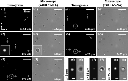
Fig. 2.
Demonstrates the sectioning ability of the lens-free tomographic microscope for 5 μm beads distributed in an approximately 50 μm-thick chamber placed at z = ∼ 0.8 mm. (A1–A5) Computed tomograms, obtained with dual-axis tomography, for various planes in the chamber volume within the range -14 μm to 25 μm. (B1–B5) Microscope images (40X, 0.65NA) for the same planes shown in A1–A5. The insets in A2 and B2 show zoomed images for a bead in the corresponding tomogram and microscope image, respectively. (A6–A8 and B6–B8) Zoomed tomograms and microscope images, in order of mention, for the region highlighted by the dashed circles in A3–A5 where two beads are axially overlapping with a center-to-center separation of approximately 20 μm. While the beads are visualized with minimal contamination due to each other in their respective planes shown in A6 and A8, the tomogram of an intermediate layer between the beads shown in A7 reveals minimal spurious details, demonstrating the sectioning ability of our lens-free tomographic microscope. Scale bars for A6–A8 and B6–B8, 5 μm. The rest of the scale bars, 50 μm.

