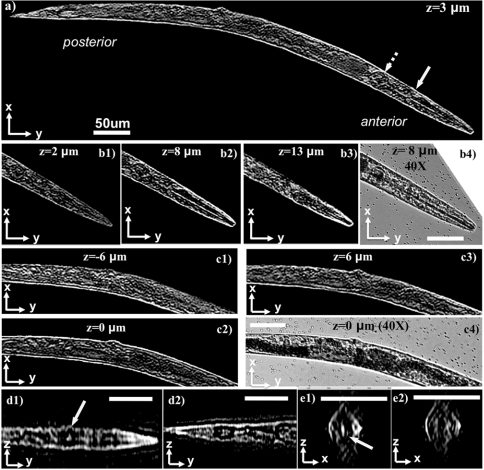
Fig. 3.
Demonstrates the application of lens-free on-chip tomography toward 3D imaging of C. elegans. (A) A tomogram for the entire worm corresponding to a plane that is 3 μm above the center of the worm. (B1–B3) Tomograms at different layers for the anterior of the worm. The pharyngeal tube of the worm, which is a long cylindrical structure with < 5 μm outer diameter, is clearly visible at z = 8 μm plane, and disappears at outer layers. (B4) A microscope image (40X, 0.65NA) for comparison. (C1–C3) Tomograms at different layers for the middle part of the worm, and a microscope image is provided in C4 for comparison. (D1 and D2) y-z ortho slices from the anterior and posterior regions of the worm, respectively. (E1 and E2) x-z ortho slices along the direction of the solid and dashed arrow in A, respectively. The 3D structure of the anterior bulb of the worm, pointed by the solid yellow arrows, can be probed by inspecting A, D1, and D3. Standard image deconvolution is applied to all the presented microscope images and tomograms to further improve their image quality as detailed in SI Text. Refer to Fig. S11 to see the raw (unfiltered) versions of these images. Movies S2 and S3 further illustrate other depth sections of the same worm. Scale bars, 50 μm.

