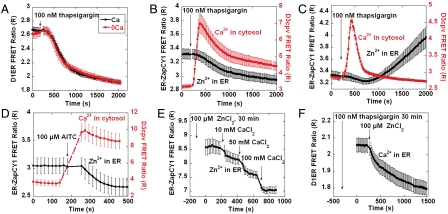
Fig. 5.
Ca2+ dynamics affect Zn2+ homeostasis in the ER. (A) Effect of thapsigargin on [Ca2+]ER. Treatment with 100 nM thapsigargin, caused [Ca2+]ER to decrease in the presence (n = 6 cells) and absence of (n = 6 cells) extracellular Ca2+. (B) Increasing [Ca2+]cyto reduced [Zn2+]ER. Treatment with 100 nM thapsigargin caused [Ca2+]cyto (n = 5 cells) to increase; [Zn2+]ER decreased (n = 6 cells) concomitantly. (C) Thapsigargin increased ER Zn2+ when extracellular calcium was absent. [Ca2+]cyto increased (n = 4 cells) and then decreased to resting level upon treatment with 100 nM thapsigargin in calcium free Hepes-buffered Hanks Balanced Salt Solution. [Zn2+]ER slightly decreased (n = 5 cells) when [Ca2+]cyto increased and then increased when [Ca2+]cyto returned to the resting level. (D) Increasing [Ca2+]cyto by activating TrpA1 reduced [Zn2+]ER. HeLa cells expressing TrpA1 were treated with AITC, causing [Ca2+]cyto (n = 4 cells) to increase and [Zn2+]ER (n = 4 cells) to decrease. (E) Effects of extracellular Ca2+ on [Zn2+]ER (n = 6 cells). HeLa cells were preincubated with 100 μM ZnCl2 for 30 min, then increasing extracellular Ca2+ reduced [Zn2+]ER. (F) Effects of extracellular Zn2+ on [Ca2+]ER (n = 5 cells). HeLa cells were treated with thapsigargin for 30 min to block the SERCA pump. Increasing extracellular Zn2+ reduced [Ca2+]ER.

