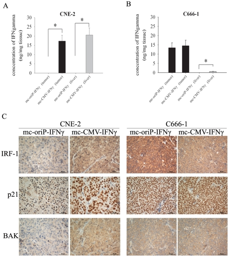
Figure 6. Tissue expression of IFNγ and immunohistochemical analysis.
(A, B) Expression level of IFNγ in tumor and liver tissue. Results are given in ng/mg of tissue. Columns, mean of three mice; bars, SD. (C) Representative immunostaining of IRF-1, p21 and BAK in CNE-2 cell- and C666-1 cell-xenografted tumors treated with mc-oriP-IFNγ or mc-CMV-IFNγ, respectively. Mice were sacrificed after three weeks of treatment, and tumors were resected and frozen for immunohistochemistry assays. IRF-1 staining shows cytoplasmic and nuclear staining, p21 staining shows nuclear staining, and BAK staining shows cytoplasmic staining. In the mc-oriP-IFNγ-treated group, intense IRF-1, p21 and BAK staining were observed only in EBV-positive C666-1 tumors. Tissue sections are shown at ×400 magnification. Scale bar represents 50 µm.

