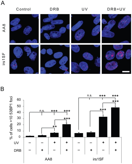
Figure 4. DRB potentiates the formation of DSBs by UV treatment, as seen by an increase in 53BP1 foci formation.
AA8 and irs1SF cells were treated with 20 µM DRB for 24 h, 10 J.m−2 UV or both. (A) Confocal through focus maximum projection images showing 53BP1 staining in red and DNA staining in blue. Bar 10 µm. (B) Quantification of 53BP1 positive cells. Cells with more than 10 bright foci were considered positive. The means and S.E. (bars) of six experiments with 200 cells counted for each experiment are shown. Values marked with asterisks are significantly different from control in T-test (**P<0.01, ***P<0.001).

