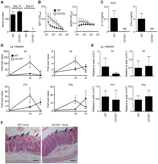
Figure 1. CD103-deficient mice are resistant to acute Trichuris infection.
WT and CD103−/− mice were orally infected with 200 Trichuris eggs. (A) Number of worms per mouse was determined microscopically at days 14 and 21 following infection. (B) Trichuris-specific IgG1 and IgG2a levels were assessed by ELISA from the sera of 21-day infected WT (•) and CD103−/− (□) mice. (C) mLN cells from WT and CD103−/− mice were restimulated with anti-CD3/CD28 Abs for 72 h and supernatants were analyzed by ELISA for production of IL-13 and IFN-γ. (D–E) Expression of Il4, Il5, Il13 and Ifng mRNA levels in the large intestine were assessed by qPCR (D) at days 14 and 21 following infection and (E) in naïve animals. Data are expressed as relative to uninfected control mice (D) and relative to actin (E). (F) Representative images of PAS-stained cecal morphology from 14-day Trichuris-infected (Tm14) WT and CD103−/− mice. Arrows indicate worms. Images were captured at 100× magnification and scale bar represents 100 µm. (A,D,E) Data are averaged from 2–3 experiments (n = 6–12); (B,C) Data is representative of 2–3 experiments (n = 8–12). ND = not detected.

