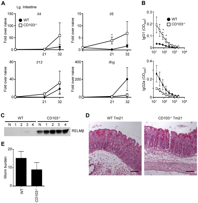
Figure 2. Chronic Trichuris infection in CD103-deficient mice.
WT and CD103−/− mice were orally infected with 25–30 Trichuris eggs. (A) Expression of Il4, Il5, Il13 and Ifng mRNA levels in the large intestine were assessed by qPCR at days 21 and 32. (B) Trichuris-specific IgG1 and IgG2a levels were assessed by ELISA from the sera of 21-day infected WT (•) and CD103−/− (□) mice. (C) Fecal samples from one uninfected (N) and four Trichuris-infected (1–4) WT and CD103−/− mice were collected on day 21 and RELMβ protein levels were assessed by Western blot. (D) Representative images of PAS-stained cecal morphology from 21-day Trichuris-infected (Tm21) WT and CD103−/− mice. Images were captured at 100× magnification and scale bar represents 100 µm. (E) Worm expulsion was assessed at day 32 following infection. (A,D) Data are averaged from 2 independent experiments (n = 6–8); (B,C) Data are representative of 2 independent experiments (n = 7–8).

