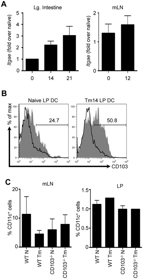
Figure 3. DC recruitment and retention in the intestine following Trichuris infection.
WT and CD103−/− mice were orally infected with 200 Trichuris eggs. (A) Itgae mRNA expression was assessed in the large intestine (left panel) and mLN (right panel) of WT mice by qPCR at days 14 and 21 following infection. (B) Representative data of CD103 expression on CD11c+ LP DCs was assessed by flow cytometry at day 14 following infection; shaded histogram: WT mice, open histogram: CD103−/− mice. (C) Frequency of CD11c+ cells in the mLN (left panel) and LP (right panel) of naïve and 14-day infected WT and CD103−/− mice. (A, and left panel of C) Data are averaged from 2–4 experiments (n = 8–16). (B, and right panel of C) Data are representative of 3–4 pooled mice from a single experiment.

