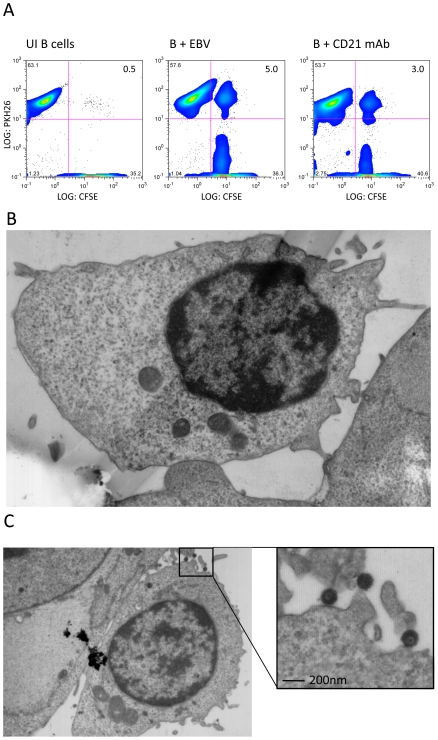
Figure 1. EBV induces firm adhesion between B cells and epithelial cells.
(A) FACS profiles of conjugate formation between CFSE-labelled primary tonsillar epithelial cells (x-axis) and PKH26-labelled primary B cells (y-axis). The B cells were uninfected, EBV-infected (MOI 100, 24 h p.i.) or incubated with agonist mAb to CD21 (BL13). Conjugates appear in the upper right quadrant and the percentages of cells are shown. (B-C) Electron micrographs of B cell-epithelial cell conjugates. EBV-infected B cells (24 h p.i.) were co-cultured with primary tonsillar epithelial cells for 1 hour and immediately fixed, embedded in Epon 812, stained with uranyl acetate and ultra-thin sectioned. (B) The sites of interaction between the cell types show firm interaction. (C) A second conjugate revealing a firm interaction between the B- and epithelial cell and the fine ultrastructure of the virus particles.

