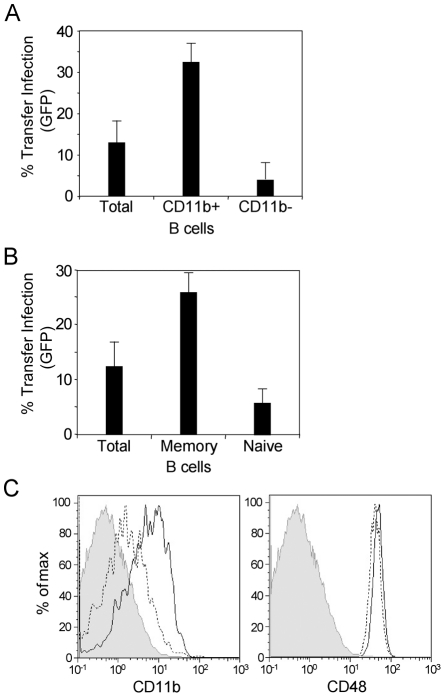
Figure 9. Transfer infection via the basolateral surface is mediated via memory B cells.
(A) Total B cells, CD11b +ve and CD11b -ve B cells were sorted and infected with EBV (24 h p.i.) and co-cultured at the basolateral surface of polarized primary tonsillar epithelial cells for 1 hour. The B cells were washed off and the epithelial cells monitored for infection by flow cytometric analysis of GFP expression after 24 hours. Infection was plotted as the mean of triplicates. (B) Total B cells, memory B cells and naïve B cells were sorted, infected with EBV, and co-cultured with the polarized epithelial cells as above. Infection was monitored by flow cytometric analysis of GFP expression and plotted as the mean of triplicates. (C) Flow cytometric analysis of naïve and memory B cell expression of CD11b and CD48. Total B cells were stained with CD27 and IgD plus CD11b or CD48. The shaded area represents the isotype control; the dotted line represents the naïve B cells (IgD+CD27−); the solid line represents the memory B cells (IgD−CD27+).

