Fig. 4.

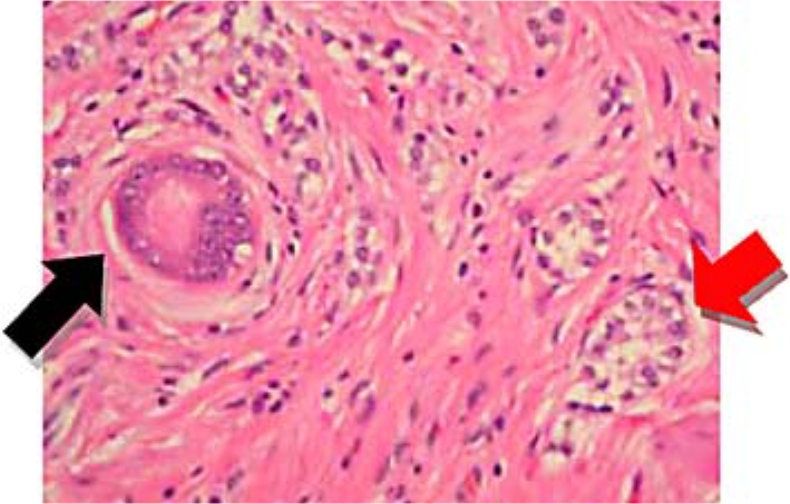
Hematoxylin and eosin staining of the polyp demonstrating glandular tissue (adenocarcinoma) (black arrow) and foci of endocrine cells (carcinoid) (red arrow).

Hematoxylin and eosin staining of the polyp demonstrating glandular tissue (adenocarcinoma) (black arrow) and foci of endocrine cells (carcinoid) (red arrow).