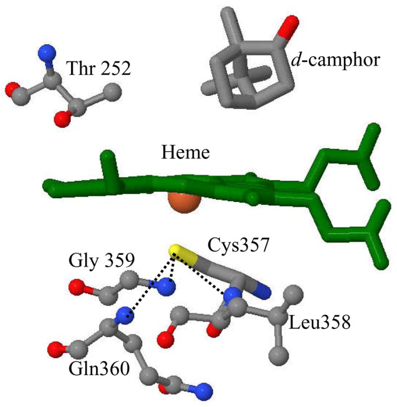
Figure 1.

The crystal structure of the camphor bound cytochrome P450cam is shown for heme, its axial ligand Cys357 and nearby amino acids (coordinates taken from the protein data bank (PDB) 2CPP72). The position of substrate, d-camphor and Thr 252 are also shown. The dotted lines represent the three hydrogen bonds between the thiolate sulfur and amide nitrogens on the proximal side.
