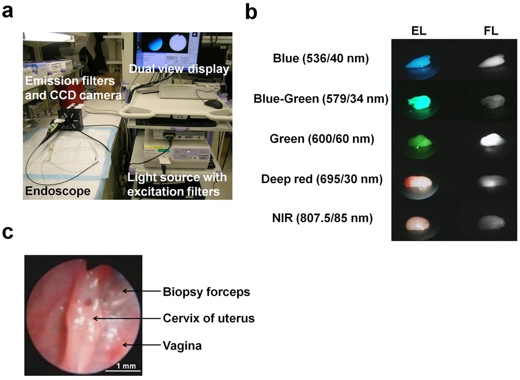
Figure 1. Multi-color fluorescence mini-endoscopic imaging of the mouse cervix.
(A) Multi-color fluorescence imaging system is based on a clinically available fiber optic endoscope and light source. Excitation light is provided by in-house designed excitation filters, which are switchable to either the visible light filter (white light) or band pass filter. Endoscopic images were obtained through a dichroic splitter, wherein the excitation light images were displayed with an image processor, and the fluorescence images were filtered by multi-color emission filters prior to reaching the EM-CCD camera. Both images are displayed side by side on the monitor. (B) Ex-vivo images of the phantoms (lymph nodes) incorporating fluorescent tracers, (Rhodamine green (close to GFP), TAMRA, RhodamineX (close to RFP), Cy5.5, ICG) showed the capability of multi-color fluorescence detection. Excitation light image (EL) and the fluorescence image (FL) were simultaneously displayed on the screen side by side. The emission filter spectrums are indicated. (C) White light endoscopic imaging of the mouse cervix. Biopsy forceps (tip = 1 mm diameter) were used to gauge the distance to the cervix which was held constant.

