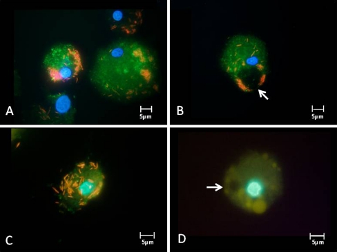
Fig. 2.
Twenty-one-day cultured sepsis-APC derived from 3 different patients (a, b, c) were incubated with red fluorescently-labeled E. coli for 3 h, followed by labeling of lysosomes with pepstatin A (green fluorescent). None of the bacteria had entered the lysosomes. Individual phagocytes show swollen lysosomes (d), here stained with lysotracker yellow. Autophagy vacuoles (arrows) appear empty

