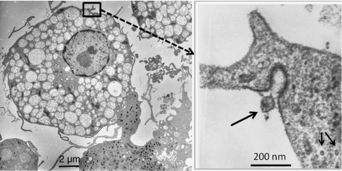
Fig. 6.
Identification of herpes virus particles (right; arrows: inserted image) in 21-day cultured sepsis-APC (left: overview) of a patient with septic shock. The extracellular virus particle (arrow) shows spiny protrusions contacting the electron dense area of the plasma membrane. Intracellular virus particles (2 arrows) lack the outer capsid structures seen in the extracellular virus particle. The cytoplasm of the APC contains multiple autophagy vacuoles containing less electron dense cytoplasm

