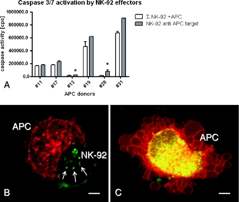
Fig. 8.
Cytolysis of cultured antigen presenting cells by NK-92 effector cells. NK-92 cells (2 × 10⁵) were co-incubated with 2 × 10⁴ cultured antigen presenting cells or 1 × 10³ APC (*) from different patients (x-axis). Caspase 3/7 activity measured by chemiluminescense (cps) after 3 h of incubation (y-axis); open columns indicate the sum of spontaneous caspase activity of NK-92 plus cultured APC (∑), grey columns indicate caspase 3/7 activity of NK-92-APC co-cultures (a). Membrane-stained NK-92- (green) and APC- (red) forming aggregates after 10 min of co-culture (b), and after 60 min intense blebbing occurs in sepsis-APC targets as well as phagocytosis of NK-92 membrane components (yellow vesicles in the sepsis-APC’s cytoplasm (red/green overlay image in c)). Bar is 5 μm

