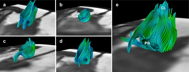
Fig. 3.
Fibre trajectories representing the anal sphincter complex in all five female subjects; 28 years, 32 years, 24 years, 27 years and 31 years of age, respectively (a–e). Not all extrapolated fibre trajectories, matching the appearance of the anal sphincter complex, were perfectly circularly orientated. This might be attributed to both the predefined fibre angle cut-off point and the potential inaccurate fibre tractography based on signal originating from various muscles and ligamentous structures converging and interweaving in this area (e.g. perineal body (PB) and coccygeal ligament (CL)) (e)

