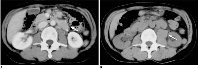
Fig. 2.
56-year-old man with paraaortic lesion.
A. Diagnostic axial contrast enhanced CT scan obtained prior to biopsy procedure with patient in supine position shows 3-cm paraaortic mass lesion (arrow). B. CT scan immediately after two punctures by posterior approach for biopsy reveals retroperitoneal hematoma (arrow).

