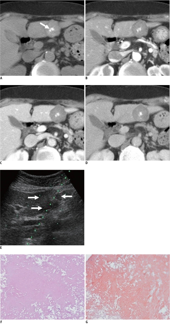
Fig. 1.
73-year-old woman with hepatic amyloidosis.
A. Unenhanced CT shows high density lobulating mass with central calcification (arrow) in segment 3 of liver. B-D. Mass shows very poor delayed contrast enhancement on arterial (B), portal venous (C) and delayed (D) phase image. E. US image during needle biopsy indicates mild heterogeneous echoic mass on segment 3 of liver (arrows). F, G. Microscopically, diffuse amyloid deposits are present without viable hepatocyte (F, Hematoxylin & Eosin staining, original magnification × 200), and Congo red stain is positive (G).

