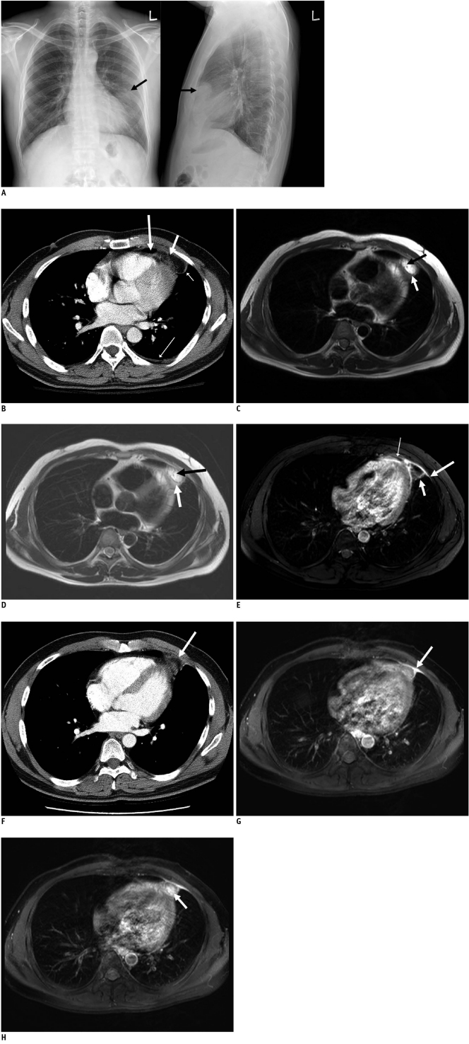
Fig. 1.
39-year-old man with sudden onset left pleuritic chest pain.
A. Chest radiographs show ovoid mass (arrows) with ill-defined margins in left anterior mediastinum. B. Enhanced CT scans show that paracardiac opacity corresponded to pericardial fat surrounded by thick rim (short thin arrow). Note associated pericardial thickening (long thick arrow), linear opacity (short thick arrow) and pleural effusion (long thin arrow). C. T1-weighted breath-hold turbo spin echo images show high signal lesion with thin hypointense rim (white arrow) and hypointense linear line (black arrow). D. T2-weighted breath-hold turbo spin echo images show high signal lesion with thin hypointense rim (white arrow) and hypointense linear line (black arrow). E. T1-weighted fat suppressed breath-hold turbo spin echo image 1 minute after gadolinium administration shows enhancement of rim (short thick arrow) and adjacent pericardium (thin arrow) and pleura (long thick arrow). F. Follow-up CT scan obtained two months after A shows that pericardial lesion has decreased in size (arrow). G. Follow-up T1-weighted fat-suppressed breath-hold turbo spin echo image obtained 1 minute after gadolinium administration and two months after image C shows peripheral rim enhancement of pericardial lesion (arrow). H. Follow-up T1-weighted fat-suppressed breath-hold turbo spin echo image obtained 5 minutes after gadolinium administration and two months after image C shows enhancement of central globular pattern (arrow) of pericardial lesion.

