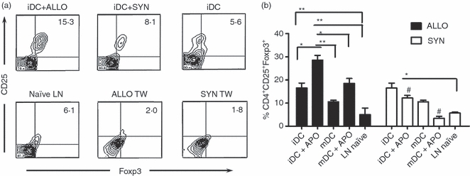
Figure 2.

Expansion of CD4+ CD25+ Foxp3+ cells after co-culture of lymph node cells (LNs) from naive mice with dendritic cells (DCs) pulsed with allogeneic or syngeneic apoptotic thymocytes (ALLO or SYN). Immature DCs (iDCs) were incubated with apoptotic thymocytes at a 1 : 5 (DC : apoptotic cell) ratio overnight. The DCs were co-cultured with syngeneic total LN cells for 5 days (1 : 5 DC : lymph node cell ratio). Cells were collected and labelled with fluorescent antibodies for CD4, CD25 and Foxp3. Dot plot (a) and bar chart (b) presentation of CD4+ CD25+ Foxp3+ cells and the same experiment in the presence of transwell chambers separating DCs and T lymphocytes (D) (gate on CD4+ cells). Lymph node cells alone and culture of iDCs with LN cells are presented as controls. Data are presented as mean ± SD and represent six independent experiments. *P < 0·05, **P < 0·005, #P < 0·005 for the comparison of both contexts.
