
Figure 3.
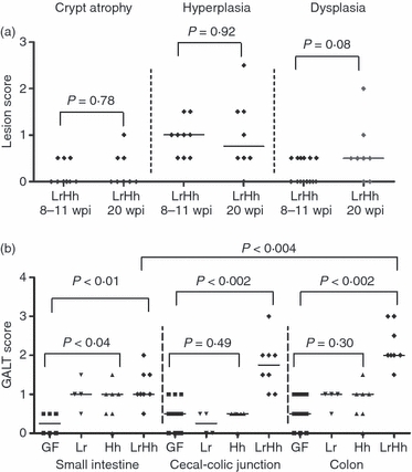
Germ-free B6.129P2-IL-10tm1Cgn (IL-10−/−) mice were colonized at 6–8 weeks of age with Lactobacillus reuteri (Lr) Helicobacter hepaticus (Hh) or L. reuteri followed in 1 week by H. hepaticus (LrHh), then killed and examined at post-mortem at 8–11 or 20 weeks post-infection (p.i.). (a) Crypt atrophy, epithelial hyperplasia and dysplasia developed only in co-infected IL-10−/− mice (LrHh); these features were absent in all other experimental groups so median scores were all significant for co-infected mice only (all P < 0.05). Differences between time-points were not significant. (b) Median gut-associated lymphoid tissue (GALT) scores for small intestine, caecal–colonic junction and the colon at 20 weeks p.i. were significantly greater in co-infected IL-10−/− mice (LrHh) compared with uninfected germ-free mice (GF) (P < 0.01, P < 0.002, P < 0.002, respectively). In co-infected mice the GALT scores were higher at the caecal–colonic junction and in representative sections of transverse and distal colon compared with small intestine (P < 0.004).