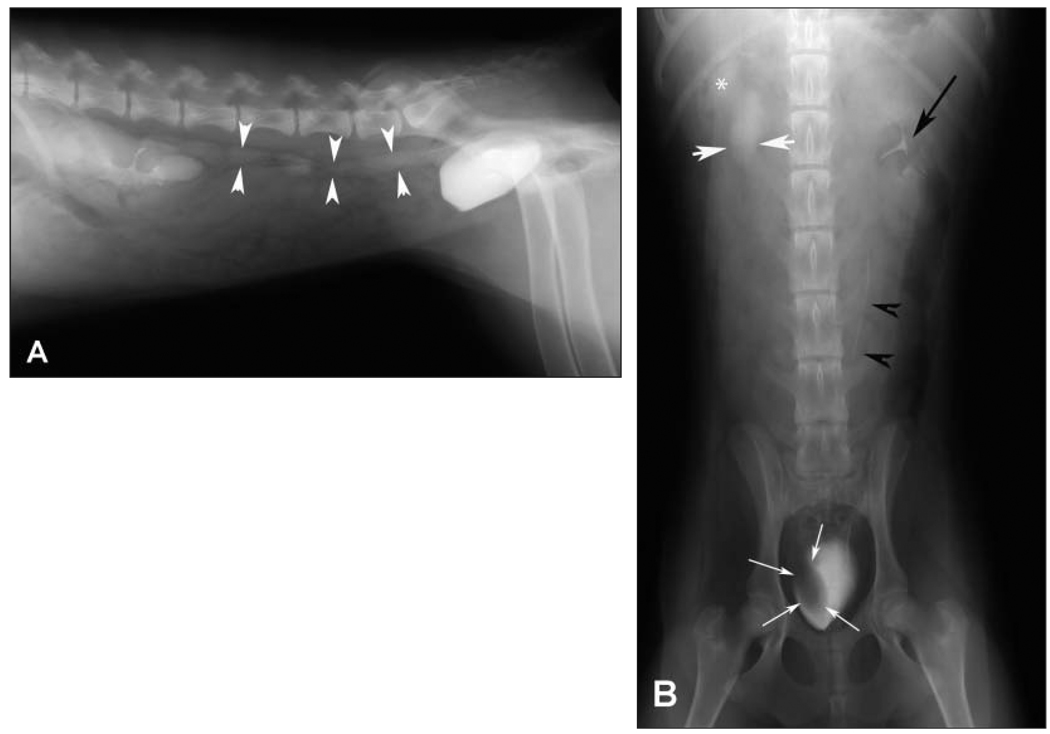
Figure 2.
Same excretory urographic images as in Figure 1. Notice that the right renal pelvis is dilated (white asterisk), the right ureter is dilated (white arrowheads), and there is a filling defect within the right side of the urinary bladder lumen (white arrows). The left renal pelvis (black arrow) and left ureter (black arrowheads) appear normal.

