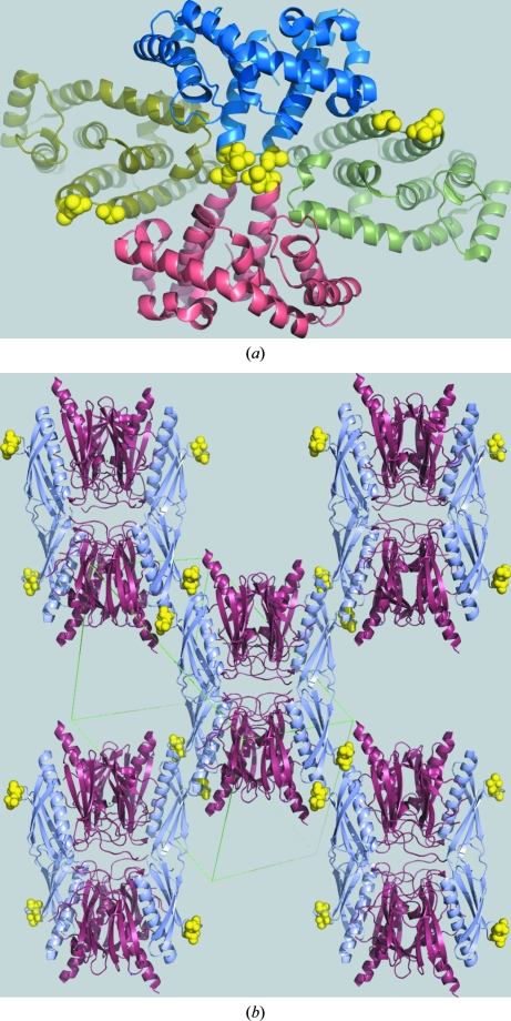
Figure 4.
Two examples of proteins crystallized by the surface-entropy reduction (SER) method. (a) The RGSL domain of the PDZRhoGEF nucleotide-exchange factor (PDB code 1htj; Longenecker, Lewis et al., 2001 ▶); the yellow spheres show the alanines introduced by mutagenesis, which mediate an isologous crystal contact across a crystallographic twofold axis. (b) The crystal structure of EpsI complexed with EpsJ (PDB code 2ret; Yanez et al., 2008 ▶); the EpsI protein (pale blue) contains two surface mutations, shown by yellow spheres, which mediate heterologous crystal contacts.

