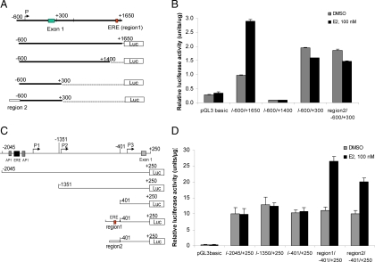
Figure 5.
Identification of the promoters and estrogen-responsive regions of the sgk3 gene. A, Schematic illustration of the 5′-flanking region as well as partial intron 1 of SGK3 transcript variant 3. The location of the P, exon 1 and the ER-binding region 1 that contains an ERE, and the structure of luciferase reporters driven by different fragments of sgk3 promoter are indicated. The nucleotides are numbered from the transcriptional start site that is assigned as +1. B, MCF-7 cells were transfected with the luciferase reporters as indicated and then cultured in the presence or absence of 100 nm E2. Twenty-four hours after transfection, luciferase activity was measured and normalized to protein concentration. Results represent three independent experiments performed in triplicates. C, Schematic illustration of the 5′-flanking region of SGK3 transcript variants 1 and 2. P1, P2, and P3 are three putative promoters as predicated by promoter predication analysis. The location of two putative AP-1 sites, one putative ERE, and exon 1, and the structure of luciferase reporters driven by different fragments of sgk3 promoter are indicated. D, The promoter activity of the sgk3 gene fragments as indicated was determined by luciferase assays. The procedures for transfection and luciferase assays are the same as described in B.

