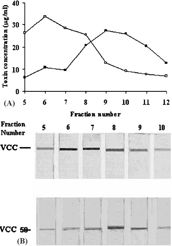
Fig. 5.

Size distribution of VCC and VCC50 micelles with rabbit erythrocyte membrane components and Triton X-100. Following incubation of the toxin with stroma at 25°C, the free toxins were removed by microfuging and the mixture was dispersed in 1 per cent Triton X-100 at 4°C. The suspension was fractionated on Sepharose CL-4B (30 × 0.7 cm) at 4° C. Fractions of 1 ml were collected and quantified for VCC (○) and VCC50 (●) by ELISA (A). Immunoblots of the fractions developed with rabbit anti-VCC antibody and goat anti-rabbit IgG conjugated to alkaline phosphatase are also shown (B).
