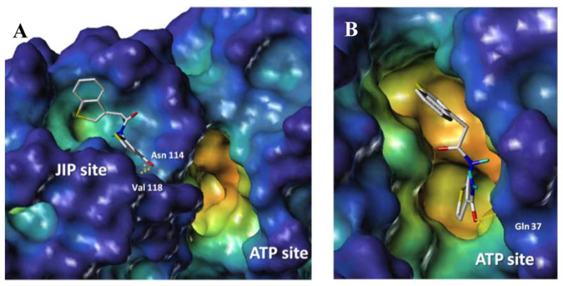
Figure 3.

Docking studies of compound 25 within the JIP (A) and ATP (B) binding sites of JNK1 (PDB ID 1UKI). The surface was generated with MOLCAD27 and color coded according to cavity depth (blue, shallow; yellow, deep). Predicted hydrogen bonding interactions are highlighted with yellow dashed lines.
