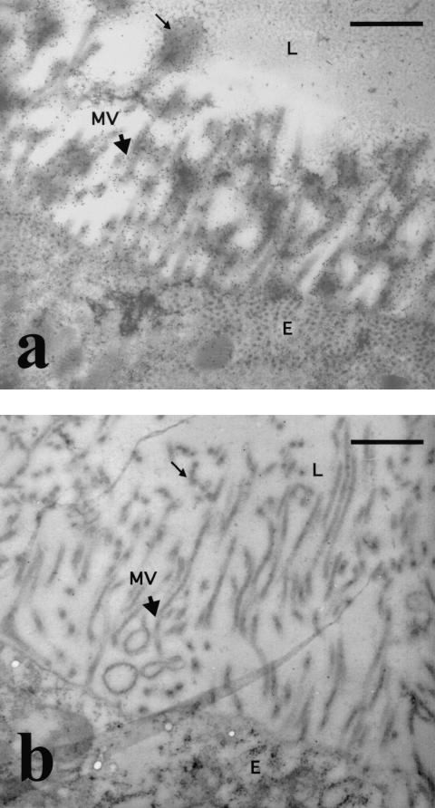
FIG. 3.
Immunoelectron microscopy of unfed An. stephensi midguts. (a) Cross-section of the midgut showing the association of MG96 with the extracellular microvilli (arrowhead) and glycocalyx (thin arrow) on the apical end of the midgut epithelial cell. (b) Cross-section of the midgut following staining with an isotype-matched control MAb. MVN, microvillus network; MV, microvilli; E, epithelial lining; L, lumens Bars, 1 μm. Magnification, ×15,000.

