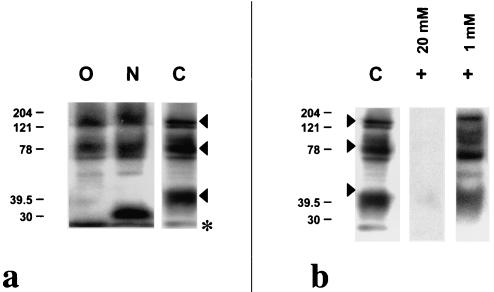
FIG. 4.
Western immunoblot analysis of An. stephensi midgut lysates following enzyme and chemical deglycosylation. (a) Lysates that were treated with 1 mU of neuraminidase plus 1 mU of O-glycosidase (lane O) or with 0.5 mU of PNGase A (lane N) were probed with MAb MG96 and compared to control lysates that were subjected to the same treatment conditions without enzyme (lane C). (b) Western immunoblot analysis of An. stephensi midgut lysates probed with MG96 following chemical deglycosylation with 1 or 20 mM sodium periodate compared to control. Molecular masses are indicated in kilodaltons at the left. Arrowheads indicate the protein doublets at ∼150, ∼80, and ∼40 kDa on untreated midgut lysate. The asterisk indicates the migration front.

