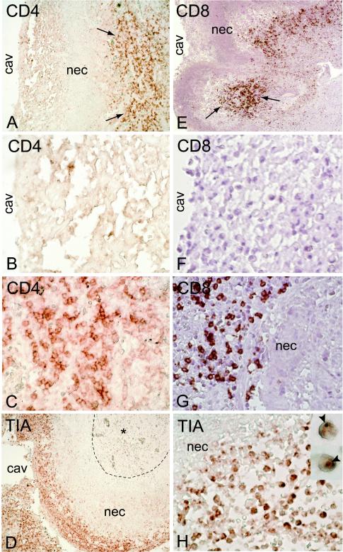
FIG. 4.
Localization of lymphoid cells in lesions of resected lungs. CD4+ (A, B, and C) and CD8+ (E, F, and G) T cells are seen in the granulomatous-fibrotic areas of the lung (arrows) but not in the necrotic (nec) zone or at the cavity (cav) surface. CD4− CD8− cytotoxic cells (TIA-1+) are seen at the cavity surface but not in the necrotic zone (D) and are less frequent in the granulomatous area (*). These cells contain cytotoxic granules that stain TIA-1+ (H, inset). Magnifications, ×4 (A, D, and E), ×40 (B, C, F, G, and H), and ×100 (H, inset).

