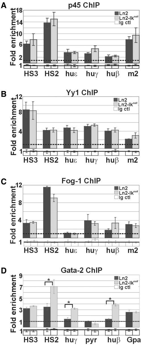
Figure 4.
Recruitment of transcriptional regulators to the huβ-globin locus in yolk sac EryC. (A–D) ChIP on ln2 and ln2-Iknull yolk sac EryC carried out with p45/Nf-e2 (p45), Yy1, Fog-1 and Gata-2 antibodies; fold enrichments (y-axis) were calculated as described in Figure 1E and are plotted as the mean ± SD of the measurements; a value of 1 (dashed line) indicates no enrichment; *P ≤ 0.05 by Student’s t-test; huε, huε-promoter; huγ, huγ-promoters; huβ, huβ-promoter; m2, mouse HS2; Gpa, Glycophorin A promoter; dark gray bars: ln2 yolk sac EryC; light gray bars: ln2-Iknull yolk sac EryC; white bars: isotype-matched Ig (Ig ctl).

