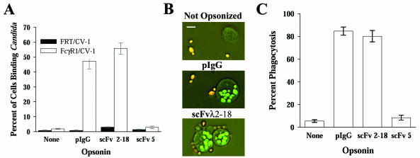
FIG. 1.
scFvλ2-18 targets C. albicans to FcR and mediates phagocytosis. (A) FcγR1-expressing epithelial cells (FcγR1/CV-1) or the parental cells (FRT/CV-1) were incubated with C. albicans strain 3153A blastoconidia opsonized as indicated. Cells that bound three or more blastoconidia were scored as positive (rosette formation). The mean percentage of cells forming rosettes ± standard error is plotted. scFv5, which does not recognize blastoconidia, was included as a mock opsonin. Anti-C. albicans pIgG- or scFvλ2-18-opsonized organisms were bound at a significantly higher level (P < 0.001 for all comparisons) than unopsonized or mock-opsonized organisms. There was no significant difference between the levels of binding of unopsonized or mock-opsonized organisms (P = 0.212). (B) Photomicrographs of THP-1 cells incubated with C. albicans strain 3153A blastoconidia, opsonized as indicated, demonstrating extracellular (orange) and intracellular (green) blastoconidia. The white bar represents 10 μm. (C) Mean percentage of THP-1 cells completing phagocytosis ± standard error of C. albicans strain 3153A blastoconidia, opsonized as indicated. The levels of phagocytosis of pIgG- or scFvλ2-18-opsonized organisms were not significantly different (P = 0.271); however, the levels of phagocytosis of either scFvλ2-18-or pIgG-opsonized organisms, compared to those of unopsonized or mock-opsonized organisms (P < 0.001 for all comparisons), were significantly different.

