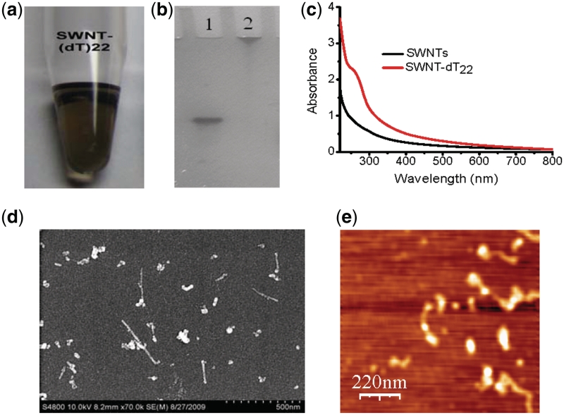
Figure 1.
Characterization of SWNTs–dT22 conjugates. (a) Photograph of the supernatant fraction obtained from the coupling steps with dT22. (b) Denaturing PAGE image of (lane 1) dT22 alone; (lane 2) purified SWNTs–dT22 conjugates. (c) UV–Vis absorption spectra of SWNTs (black line) and SWNTs–dT22 conjugates (red line); (d) SEM image of SWNTs–dT22 conjugates. After the sample was purified as described in ‘Materials and Methods’ section, SEM shows that SWNTs are dispersed upon DNA conjugation. (e) AFM image of SWNTs–dT22 conjugates. AFM indicates that dT22 are conjugated to SWNTs and the formed conjugates are dispersed.

