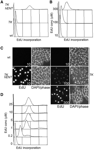
Figure 1.
EdU incorporation in fission yeast strains expressing TK and hENT1. (A) Wild-type (P2), TK (adh1:tk, P2471) and TK hENT1 (adh1:tk adh1:hENT1, P2470) expressing strains were grown in YE3S containing 10 µM EdU for 3 h then fixed and conjugated to Alexa Fluor 488 azide before analysis by flow cytometry, (B) adh1:tk (P2471) cells were grown in YE3S containing the indicated concentrations of EdU for 3 h then analysed as in (A), (C) Cells from (A) and (B) were imaged by fluorescence microscopy. Bar = 10 µm and (D) adh1:tk adh1:hENT1 (P2470) cells were grown in YE3S containing the indicated concentrations of EdU for 3 h then processed as in (A) and analysed by flow cytometry. Linear x-axes are used to show EdU incorporation in all panels in this figure.

