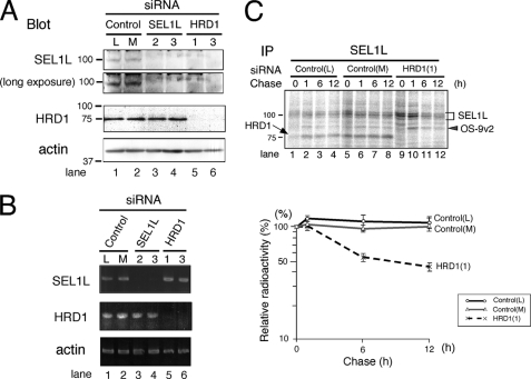
FIGURE 1.
SEL1L is destabilized by HRD1 knockdown. A, expression of SEL1L and HRD1 in HEK293 cells treated with siRNA targeted for SEL1L or HRD1 is shown. HEK293 cells were treated with 30 nm of StealthTM siRNA for 48 h. As a negative control, StealthTM siRNA Negative Control Low GC (L) and Medium GC (M) were used. Twenty μg of cell lysates were separated by 10% SDS-PAGE. The expression of each protein was analyzed by Western blotting. Mouse monoclonal anti-SEL1L antibody was used to detect SEL1L (upper panel). The positions of the molecular weight standards are shown on the left. B, detection of SEL1L and HRD1 transcripts by RT-PCR is shown. One mg of total RNA prepared from HEK293 cells treated with siRNA as in A was used. C, pulse-chase analysis of endogenous SEL1L is shown. HEK293 cells treated with siRNA (as in A) were labeled for 20 min and then chased for the time indicated. Cells were extracted in a buffer containing 3% digitonin. The positions of SEL1L, OS-9v2, and HRD1 are indicated by a bracket, gray arrowhead, and arrow, respectively. The positions of the molecular weight standards are indicated on the left. The relative radioactivity of SEL1L was quantified, and the signal intensity at the end of pulse-labeling period was set as 100% (lower panel). Data shown are the mean and corresponding S.E. of three independent experiments. IP, immunoprecipitate.

