FIGURE 8.
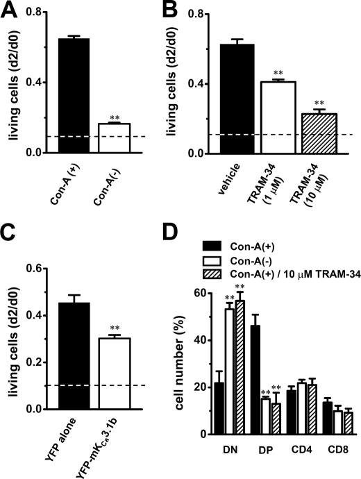
Effects of mKCa3.1b overexpression and IKCa current blockade on cell growth and CD4/CD8 phenotype expression in mouse thymocytes. Cell viability at d2 was evaluated by MTT cell proliferation assay. Value of living cells was expressed as the absorption spectra of the formed formazans and was calculated relative to that in thymocytes at d0. A, value of living cells in Con-A(−) thymocytes (open column) at d2 was compared with that in Con-A(+) thymocytes (solid column) (n = 6 for each). **, p < 0.01 versus Con-A(+). B, value of living cells in Con-A(+) thymocytes treated with 1 μm (open column) and 10 μm (hatched column) TRAM-34 at d2 was compared with that in Con-A(+) thymocytes treated with vehicle (0.1% DMSO) (n = 6 for each). **, p < 0.01 versus vehicle. C, value of living cells in Con-A(+) thymocytes transfected with YFP-mKCa3.1b (open column) at d2 was compared with that in Con-A(+) transfected with YFP alone (solid column) (n = 4 for each). **, p < 0.01 versus YFP alone. Dashed lines show value of living cells in thymocytes cultured in the absence of Con-A. D, reduced numbers of CD4(+)CD8(+) double-positive (DP) thymocyte populations in Con-A(−) and Con-A(+) treated with TRAM-34. Thymocytes were stained for CD4 and CD8 and examined by flow cytometry. The bar graph summarized each subset from separate (n = 5 for each) experiments as follows: CD4(−)CD8(−) double-negative (DN), CD4(+)CD8(+) double-positive (DP), CD4(+)CD8(−) single-positive (CD4), and CD4(−)CD8(+) single-positive (CD8). **, p < 0.01 versus Con-A(+).

