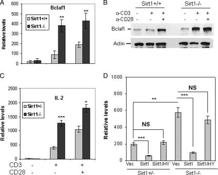
FIGURE 1.
Sirt1 inhibits Bclaf1 expression in T cells. A and B, naive CD4+ T cells from Sirt1+/+ and Sirt1−/− mice were stimulated with anti-CD3 or CD3 plus anti-CD28 antibodies for 16 h. Total RNA was isolated, and the levels of Bclaf1 (A) and IL-2 (B) were determined by real-time PCR. C, primary T cells were stimulated with anti-CD3 or with anti-CD3 plus anti-CD28 for 24 h. Bclaf1 protein expression in the stimulated cells was analyzed by Western blotting using anti-Bclaf1 (top panel) antibody. The same membrane was reblotted with anti-actin as a loading control (bottom panel). D, Sirt1+/+ and Sirt1−/− MEF cells were transfected with Sirt1 or Sirt1/HY mutant. The mRNA expression levels of endogenous Bclaf1 in the transfected cells were analyzed by real-time PCR using β-actin mRNA as an internal control. vec, vector. Student's t test was used for statistic analysis, NS, no significant difference. Error bars indicate mean ± S.D.; *, p < 0.05; **, p < 0.01; ***, p < 0.005.

