FIGURE 6.
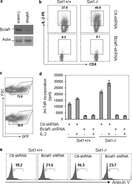
Bclaf1 knockdown inhibits Sirt1-null T cell activation. A, mouse primary T cells were isolated from Sirt1+/+ and Sirt1−/− mice and infected with lentivirus that carries shRNA specific to Bclaf1 or a control shRNA. GFP-positive cells were sorted 2 days after infection. Bclaf1 protein expression in sorted cells was determined by Western blotting with anti-Bclaf1 antibody (top panel). Protein expression of actin was analyzed as a loading control (bottom panel). B and C, mouse primary CD4+ T cells from Sirt1+/+ and Sirt1−/− mice were infected with lentivirus that carries shRNA specific to Bclaf1 or a control (Ctl) shRNA. The production of IL-2 and cell surface CD4 expression in gated GFP+ cells was analyzed by intracellular staining followed by flow cytometry (B), and the percentages of GFP+ cells at 2 days after infection are shown (C). D, GFP+ cells were sorted and restimulated with anti-CD3 (1 μg/ml) or anti-CD3 plus IL-2 (1 ng/ml). Proliferation was analyzed by [3H]thymidine incorporation (3H-TdR). Error bars indicate mean ± S.D. E, GFP+ cells were gated, and apoptotic cells were analyzed by annexin V staining. FSC, forward scatter; 3H-TdR, [3H]thymidine.

