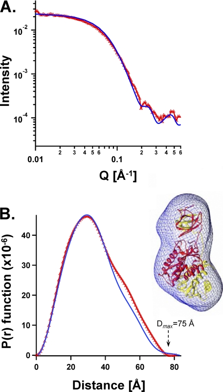
FIGURE 2.
Comparison of the crystal structure with solution SAXS dimensions and shapes of the same NTD-lacking two-domain ASV IN fragment (IN(49–286) F199K at 1.1 mg/ml). A, experimentally determined SAXS scattering is shown in red triangles. Values calculated from the crystal structure of the same IN fragment (PDB coordinate file 1C1A) using the Crysol program are represented by a blue line. B, experimentally derived plot of P(r) function for the SAXS data is compared with values calculated from the same crystal structure. Color code is the same as in A. Right, SAXS envelope shape derived from the experimental data is portrayed as a blue wire mesh, and the atomic resolution coordinates of 1C1A are shown within the SAXS envelope, with one monomer colored red and the other yellow.

