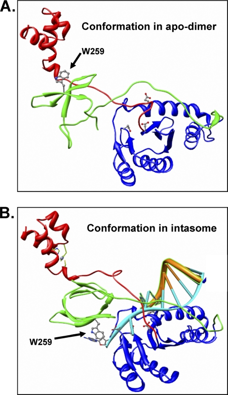
FIGURE 8.
Conformational change required for the transition from a reaching dimer to the intasome complex with DNA. A, conformation of a single ASV IN subunit in the apo-IN reaching dimer. B, conformation predicted from the inner dimer of an intasome complex that includes the viral substrate DNA. The subunit structure is modeled from the PFV intasome (PDB code 3OYA (38)). The change in orientation of the CTD residue Trp-259, shown in ball-and-stick, is highlighted with arrows. Active site residues are also shown in ball-and-stick fashion. A supplemental movie that simulates the conformational change between these two states is provided.

