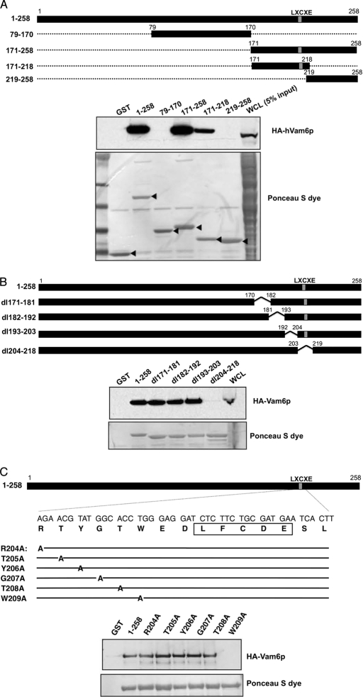
FIGURE 3.
Mapping the hVam6p binding site on MCV LT antigen. A, diagram of GST-tagged LT deletion constructs. GST-LT fusion proteins were incubated with protein extracts of 293FT cells transfected with HA-hVam6p. Bound proteins were revealed by immunoblotting with anti-HA antibody (top panel). The GST fusion LT proteins were visualized by Ponceau S staining (bottom panel). B, further deletion analysis of the 171–218 subregion in the context of 1–258 LT. Ponceau S shows comparable expression of LT constructs and 1–258 has appropriately retarded migration (bottom panel). Co-immunoprecipitation shows that only dl204–218 loses the ability to interact with hVam6p (top panel). C, schematic diagram of MCV LT alanine substitution mutants based on localization of hVam6p binding to residues 204–218 (A and B). LFCDE, Rb-binding domain.

