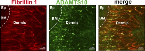
FIGURE 3.
Co-localization of ADAMTS10 with fibrillin-1 in human skin. A, immunofluorescent visualization of the distribution of fibrillin-1 (red) and ADAMTS10 (green) in human skin. The locations of the epidermis (Ep), dermis, and basement membrane (BM; broken white line) are shown. Note the substantial overlap between ADAMTS10 and fibrillin microfibrils in the papillary dermis (closest to the basement membrane) with relatively lower ADAMTS10 staining intensity in the further removed reticular dermis (center and right of each panel). Scale bar, 10 μm. Negative controls indicating antibody specificity are shown in the supplemental data.

