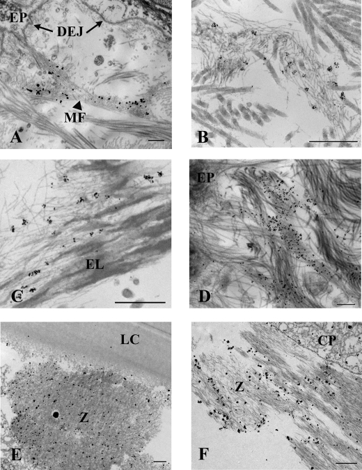
FIGURE 4.
Immunoelectron microscopy illustrates that ADAMTS10 is specifically bound to tissue microfibrils in skin and zonules. ADAMTS10 was localized en bloc using mAb 287-3E2 followed by 1-nm gold secondary conjugate and then gold-enhanced to allow visualization at low magnification. Labeling of microfibril bundles (MF) is most intense close to the dermal-epidermal junction; however, labeling is absent immediately adjacent to the epithelium (EP) and the basement membrane at the dermal-epidermal junction (DEJ; A, arrows). Immunogold particles seen at higher magnification following slightly less time in gold enhancement solution demonstrate that ADAMTS10 localizes to microfibrils in small clumps represented by a cluster of gold particles (B; image taken near the dermal-epidermal junction) also seen among elastin-associated microfibrils in the shallow reticular dermis (C). An image collected at 0° tilt through an exceptionally thick section (300 nm; D) of immunolabeled skin shows several labeled microfibril bundles intersecting the lamina densa of the dermal-epidermal junction best appreciated in the aligned tilt series (supplemental Video File 2). Ciliary zonules (Z) intersecting both the lens capsule (LC; E) and ciliary process (CP; F) are well labeled with antibody to ADAMTS10. Scale bars for A–D, 500 nm; scale bars for E and F, 1 μm.

