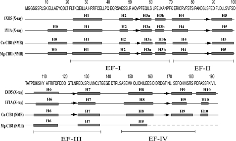
FIGURE 1.
Secondary structure arrangements of the two available x-ray crystal structures of Ca2+-CIB1 (1XO5 and 1Y1A) and the NMR solution structures of Ca2+-CIB1 and Mg2+-CIB1. The positions of the four EF-hand helix-loop-helix structures are indicated. Boxes indicate α-helices, and arrows indicate β-strands.

