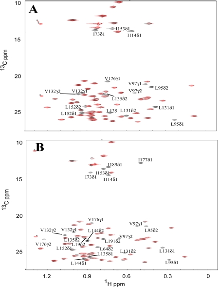
FIGURE 5.
Superimposed 13C-HSQC spectra of the Ile, Leu, and Val methyl groups of CIB1 and CIB1 bound to αIIb in the absence (black) and in the presence of 6 eq of TEMPOL (red). The same amount of TEMPOL (6 eq) was added into 12C,2H,15N-uniformly and Ile-δ1-13CH3,Leu,Val-13CH3,12CD3-labeled Ca2+-CIB1 (A) and Mg2+-CIB1 (B) samples at the same concentration (0.3 mm). The peaks marked with an asterisk are most likely from the 13C isotopic natural abundance of TEMPOL as those peaks do not appear for the protein alone.

