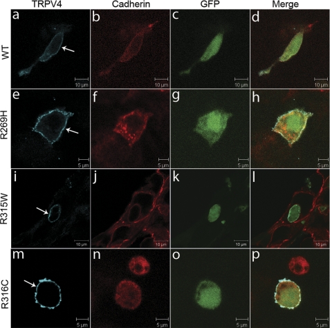
FIGURE 2.
Physiological localization of wild-type and mutant TRPV4 on the plasma membrane. Confocal microscopy was performed using HEK293 cells transfected with plasmids pIRES2-ZsGreen1 containing wtTRPV4 (a–d), TRPV4R269H (e–h), TRPV4R315W (i–l), and TRPV4R316C (m–p). Cells expressing exogenous TRPV4 were labeled by ZsGreen1 GFP (c, g, k, and o). TRPV4 is shown by blue (a, e, i, and m) and cadherin by red (b, f, j, and n). Merged images are shown on the right panels (d, h, l, and p). Arrows indicate TRPV4 signals on the plasma membrane. Representative images are provided. For each condition, at least 50 cells were analyzed in more than two independent experiments.

