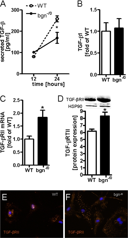
FIGURE 4.
Increased expression of TGF-βRII in bgn−/0 fibroblasts. A, TGF-β ELISA revealed lower levels of secreted TGF-β in medium conditioned by bgn−/0 fibroblasts; n = 3. B, mRNA expression of TGF-β1 was not different between the two genotypes; n = 6. C, mRNA expression of TGF-βRII was significantly elevated in bgn−/0 fibroblasts. D, immunoblotting of TGF-βRII; n = 5. E and F, immunocytochemical detection of TGF-βRII in primary cardiac fibroblasts in the first passage, representative images are shown of n = 3. The staining revealed increased TGF-βRII expression in bgn−/0 fibroblasts. Analysis was performed 24 h after plating in 5% FCS. Data are presented as mean ± S.E.; *, p < 0.05.

