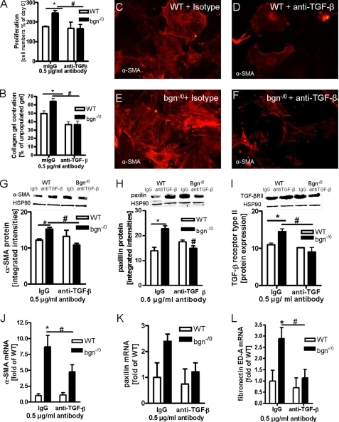
FIGURE 5.
Reversion of myofibroblast phenotype by neutralizing endogenous TGF-β. A, cell growth in the presence of either non-immune mouse IgG (mIgG) as isotype control or neutralizing antibody to TGF-β -1, -2, -3 (anti-TGF-β) after 24 h in 5% FCS. Both antibodies were used at 0.5 μg/ml. Anti-TGF-β significantly inhibited proliferation of bgn−/0 fibroblasts; n = 3. B, OSCR assay in the presence of mIgG or anti-TGF-β. Anti-TGFβ blocked enhanced collagen gel contraction by bgn−/0 fibroblasts (black bars); n = 3. C–F, immunocytochemical staining of α-α-SMA of WT (C and D) and bgn−/0 fibroblasts (E and F) treated with either mIgG (C and E) or anti-TGF-β neutralizing antibody (D and F). Anti-TGF-β reduced α-SMA-positive stress fibers in bgn−/0 fibroblasts; 200× magnification; representative images of n = 3 experiments. G, immunoblotting confirmed the responsiveness of α-SMA to anti-TGFβ antibodies; n = 3. H and I, protein expression as evidenced by immunoblotting of paxillin (H) and TGF-βRII (I) were down-regulated by neutralization of TGF-β in bgn−/0 fibroblasts as well. HSP90 served as loading control; n = 3. J–L, mRNA expression of α-SMA, paxillin, and the fibronectin fragment ED-A were analyzed by quantitative real-time PCR. α-SMA and fibronectin ED-A mRNA expression were increased in bgn−/0 fibroblasts and were decreased by neutralizing TGF-β. Paxillin mRNA showed a similar trend; n = 4. Experiments were performed 24 h after plating in 5% FCS Data are presented as mean ± S.E.; *,# p < 0.05.

