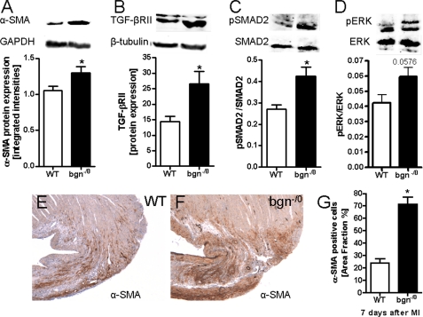
FIGURE 8.
TGF-βRII protein expression and SMAD2 phosphorylation were increased post-MI in bgn−/0 mice. Immunoblotting using left ventricular tissue extracts 3 days post-MI. A, Western blotting of α-SMA in tissue extracts from infarcted ventricles confirmed increased α-SMA expression in bgn−/0 mice. B, TGF-βRII protein expression. C, increased ratio of phosphorylated SMAD2 to total SMAD2 in bgn−/0 hearts. D, ratio of phosphorylated ERK to total ERK1/2 revealed only a trend toward increased phosphorylation of ERK1/2; in A–D, n = 5. E and F, immunostaining of α-SMA 7 days after experimental MI. Shown are representative images of the peri-infarct zone confirming up-regulated α-SMA expression in bgn−/0 mice; n = 4. G, quantification of α-SMA immunohistochemistry as in D and E was performed using ImageJ (NIH). Data are expressed as mean ± S.E.; *, p < 0.05.

