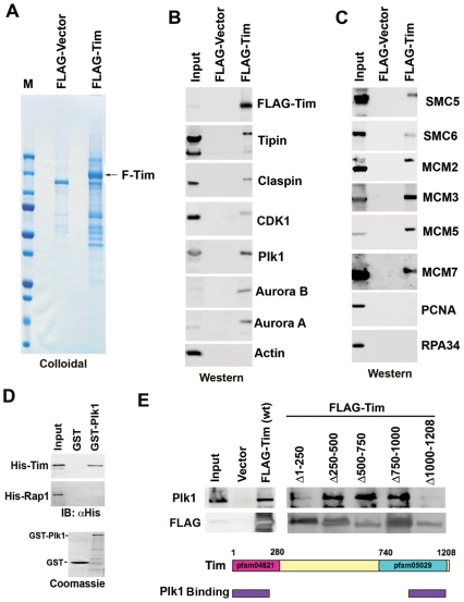
Figure 1. Proteomic analysis of FLAG-Tim complex.
A) FLAG-Tim expressing 293 cell lines was used to purify Tim complex using FLAG-agarose. FLAG-vector control or FLAG-Tim proteins were visualized by SDS-PAGE and colloidal blue staining. B) Western blot analysis of FLAG purified proteins from FLAG-Vector or FLAG-Tim expressing cells. Input represents 5% of the starting material from the FLAG-Tim stable cell line. Antibodies for FLAG, Tipin, Claspin, CDK1, PLK1, Aurora B1, Aurora A1, or Actin are indicated. C) Same as in B, except with antibodies to SMC5, SMC6, MCM2, MCM3, MCM5, MCM7, PCNA, and RPA34. D) Baculovirus expressed His-Tim or His-Rap1 were assayed for binding to purified GST or GST-Plk1 by GST-pull down assay. Input and bound proteins were analyzed by Western immunoblot (IB) with anti-His antibody. GST-fusion proteins were visualized by Coomassie staining of SDS-PAGE gels (lower panel). E) Western blot of immunoprecipitates with cells transfected with FLAG-vector, FLAG-Tim wt, or FLAG-Tim deletion mutants (as indicated above each lane). Input is indicated. IPs were assayed by Western blot for Plk1 (top panel) or FLAG (lower panel). Schematic of Tim protein showing that amino and carboxy-terminal conserved domains as pfam04821 and pfam05029, and summary of Plk1 binding is indicated with purple rectangles.

